Plug FerryLive's dental algorithms
into your system
Six modalities: intraoral, panoramic, cephalometric, facial, CBCT and IOS. On-premises deployment or API access, structured output for everything, data never leaves your campus. On the right is a replay of a real inference run.
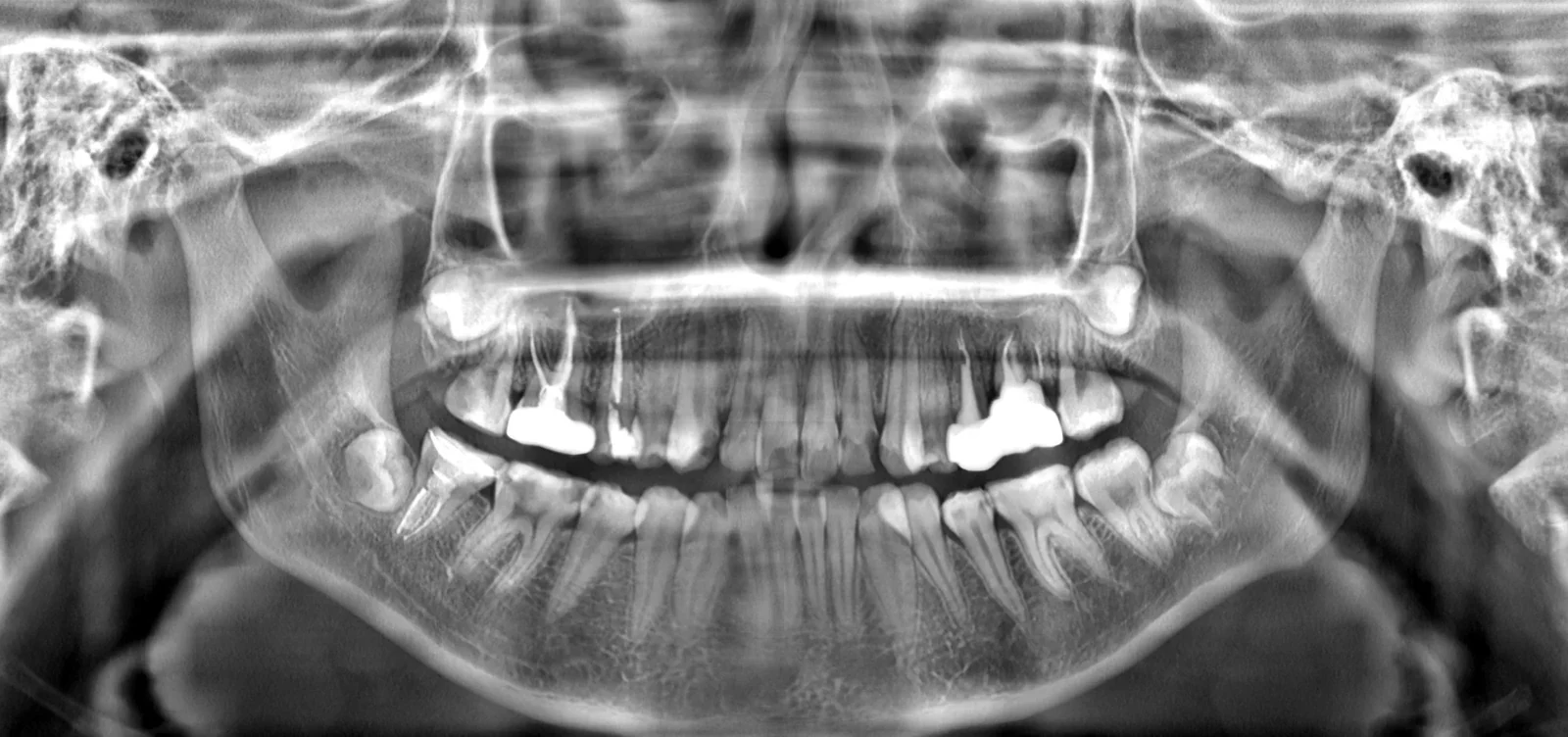
Public demo panoramic · annotations are raw algorithm output, not retouched · clinical use requires doctor confirmation
An image goes in, structured results come out
Every annotated image below is the raw output of FerryLive AI Engine on this site's demo data, and every 3D model is a real segmentation result.
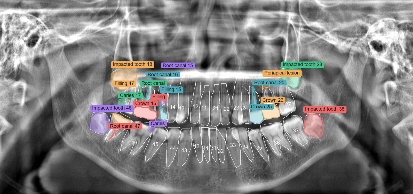 PanoramicFDI + PanoramicLesion
Panoramic analysis
One panoramic X-ray gives tooth segmentation and lesion detection in a single pass, suited to batch pre-screening and workstation integration.
- Contour-level instance segmentation with a polygon and bounding box per tooth
- Caries, impacted teeth, periapical lesions, alveolar bone loss, fillings, root canal fillings, restorations and more
- Results carry class, confidence and pixel-aligned coordinates
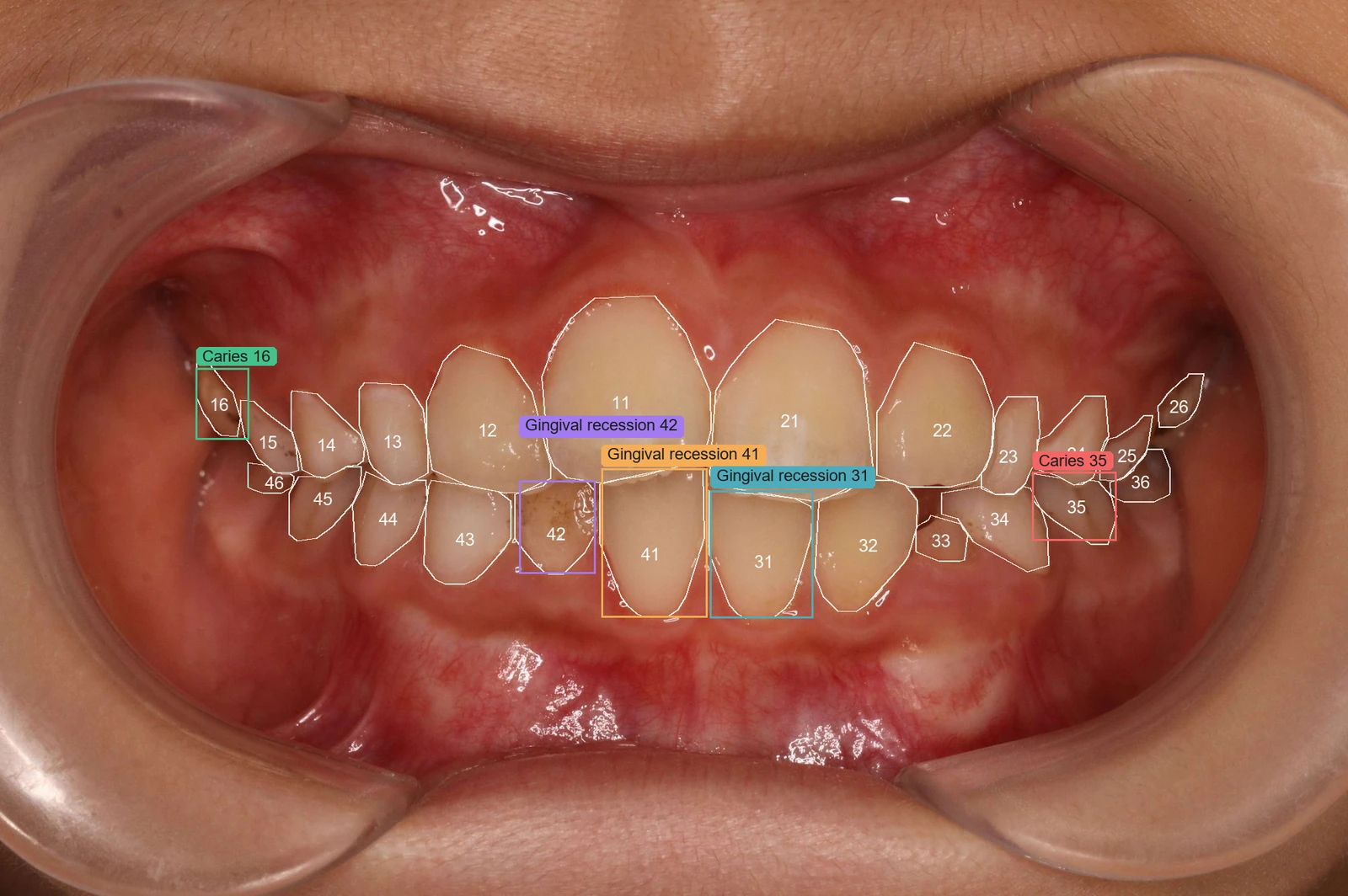 52 FDI classes + 6 lesions + 4 image-level
Intraoral analysis
Tooth-level models locate lesions tooth by tooth while an image-level model gives overall conclusions, all with boxes and confidence.
- Tooth level: caries, tooth wear, wedge defects, residual crowns, gingival recession, fluorosis
- Image level: crowding, calculus, gingival abnormality, discoloration
- Lesions are assigned to teeth by IoU, ready to drop into a record
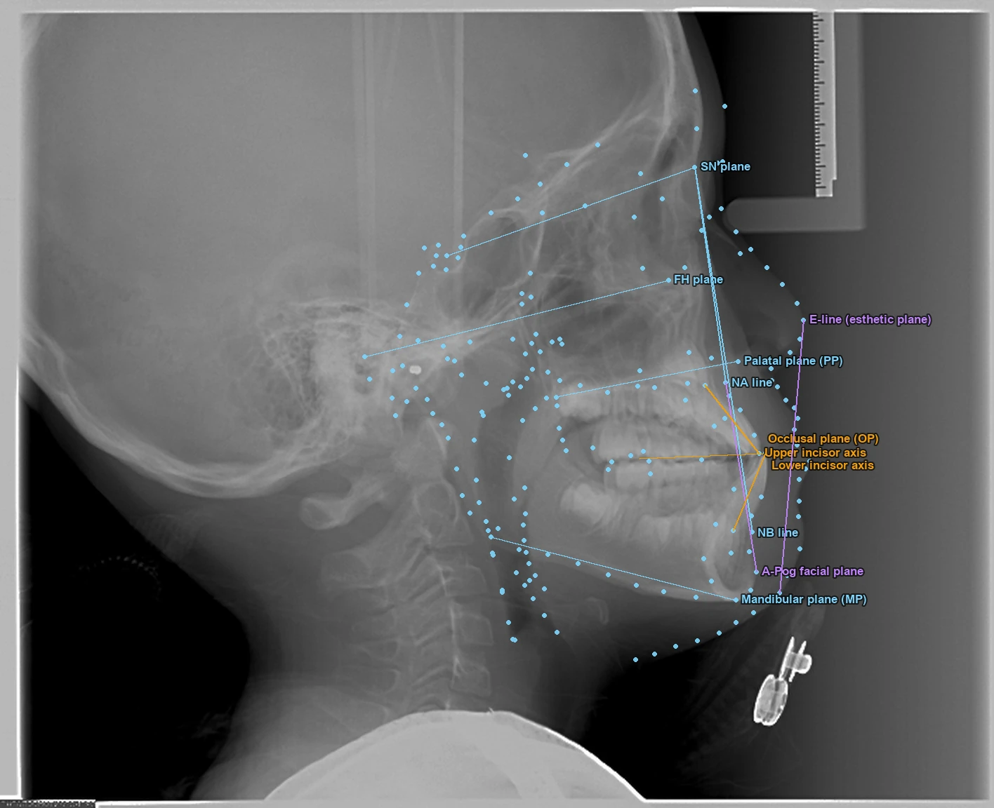 CeLDA+ · 201 points · 26 measurements
Cephalometric analysis
Landmarks are placed automatically on lateral cephalograms, then sagittal, vertical, dental and soft-tissue metrics are computed with reference planes drawn in.
- Basic 46-point model; enhanced 201 points + 26 measurements
- SN, FH, palatal, occlusal, mandibular and esthetic planes drawn automatically
- Every metric shows its reference range and a high/low flag for quick review
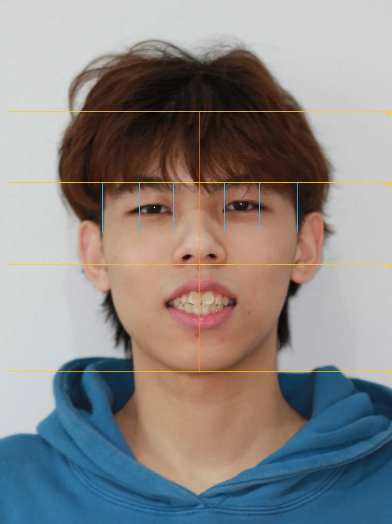 FaceLandmarker 478 points + esthetics
Facial and smile esthetics
Facial thirds and fifths, nasolabial angle, buccal corridor and midline deviation, with frontal, oblique and profile paths chosen automatically from the shooting angle.
- Thirds, fifths and lower-face ratios compared against ideal values
- Smile esthetics: gingival display grade, buccal corridor ratio, smile index, commissure tilt
- Profile path adds nasolabial angle and facial convexity
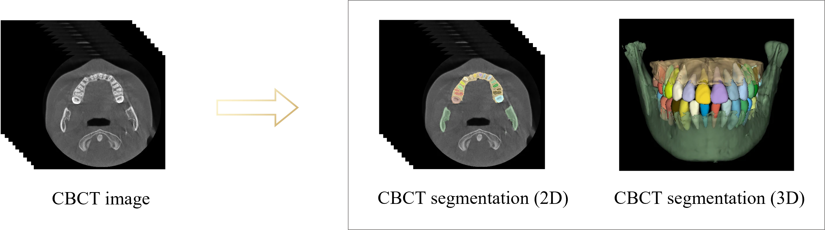
CBCT tooth and jaw segmentation
A hierarchical two-stage network segments every tooth and both jaws from the 3D volume, handling missing teeth, metal artifacts and malocclusion. The model on the left is real output, colored per tooth.
- ROI extraction, centroid and skeleton prediction, multi-task tooth segmentation, cascaded bone segmentation
- Outputs per-tooth 3D meshes and MPR view data
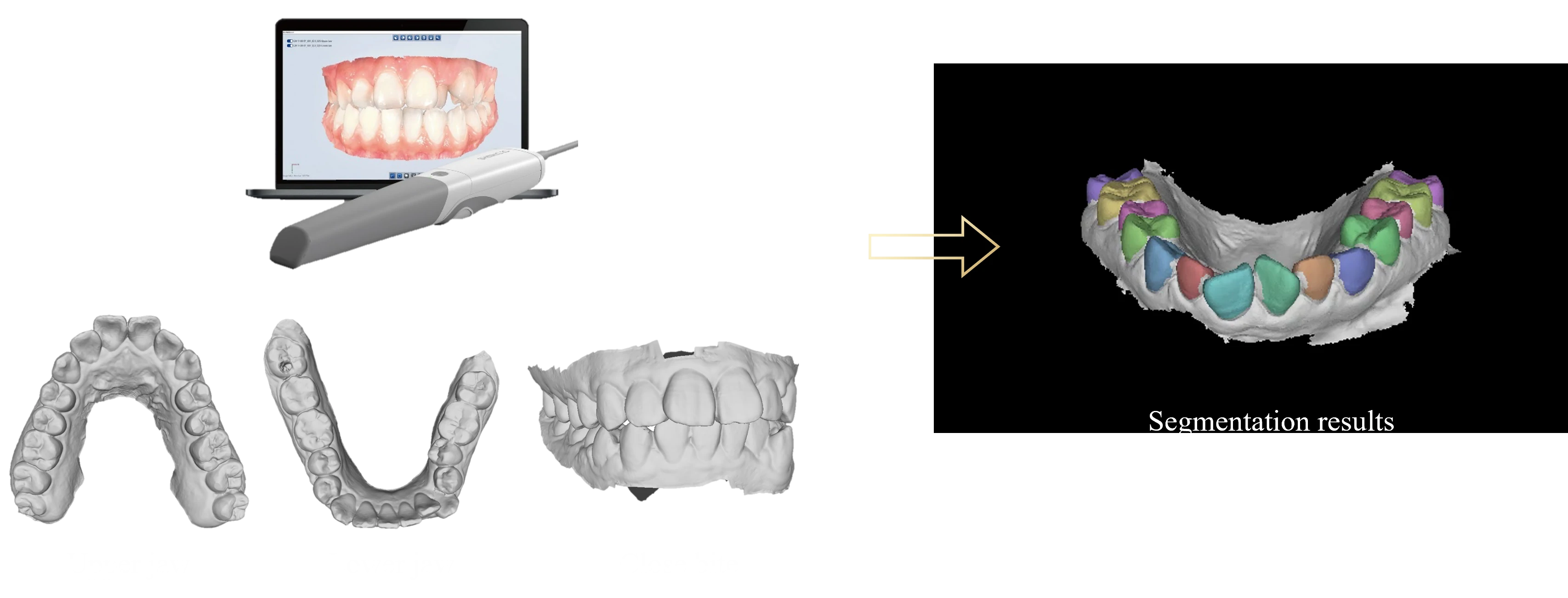
IOS per-tooth segmentation
A centroid-guided network segments and numbers every tooth in an intraoral scan, robust to crowding, missing teeth, blurred boundaries and unusual shapes.
- Locate tooth centroids first, then segment and number each tooth
- Results feed setup prediction, periodontal measurement and crown design directly
Setup prediction, from scan to after treatment
A diffusion model arranges the teeth from an intraoral scan and outputs an interactive post-treatment arch. On the left is a real result: drag the slider and every tooth moves from its initial pose to the predicted one using the algorithm's own translation and rotation.
- Automatic alignment network validated on 800 pre/post treatment cases
- Arch, neighbor and single-tooth constraints keep movements physically plausible
- Outputs a standard GLB plus per-tooth transform matrices for any viewer
Simulations support doctor-patient communication and are not a diagnosis.
One smile photo, see the result first
A smiling photo goes through three-stage alignment to produce the post-treatment smile in about 30 seconds, the lowest-effort way to let patients preview. Drag to compare.
- Only a smile photo is needed, no scan
- For communication and case acceptance, not diagnosis


Drag the slider to compare · real model output
CBCT registered to IOS, six sites measured automatically
The heatmap and slice below come from the same real case: probing depth at 168 sites on 28 teeth measured by the algorithm along each tooth's long axis. Click any tooth for its six-site values.
Full-mouth periodontal heatmap · probing depth PD
IOS–CBCT registration passed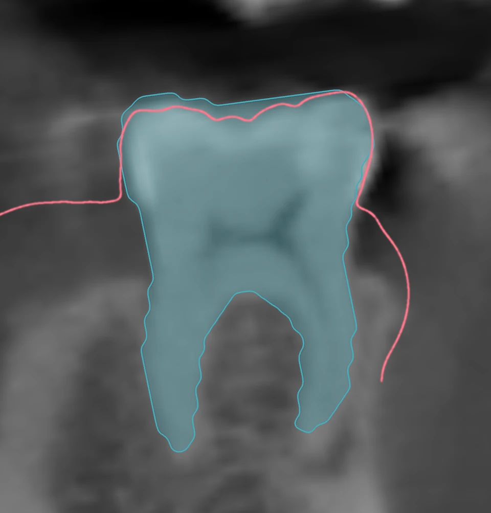
- Validated on 83 paired cases (about 2,264 teeth), mean error 0.031 mm
- Long-axis measurement cuts error by roughly an order of magnitude versus shortest-distance methods
- Outputs six-site PD / GM / CAL with automatic staging and grading under the 2018 EFP/AAP classification
- Registration quality is reported in three grades; mismatched data is flagged instead of forced
Nine areas, one engine
Every capability comes from models running in FerryLive's clinical products. Published work lists the venue and validation data; unpublished work lists only what is implemented.
Intraoral analysis
Per-tooth instance segmentation, lesion detection and image-level conclusions.
- 52 FDI classes including primary teeth
- Caries, wear, wedge defects, residual crowns, recession, fluorosis
Panoramic analysis
Tooth segmentation and 20-class lesion detection in one pass.
- 48-class contour-level instance segmentation
- Boxes, contours, classes and confidence out
CBCT tooth and jaw segmentation
Two-stage network for per-tooth and jaw segmentation.
- 4,215 cases, 15+ centers
- AI ~5 min versus 150 min manual
IOS per-tooth segmentation
Centroid-guided point-cloud segmentation and numbering.
- 1,000 patients, 2,000 arches
- PLY / STL in, FDI-colored mesh out
Cephalometric and facial analysis
Ceph landmarks plus facial esthetic metrics.
- Basic 46 points, enhanced 201 points + 26 measurements
- Facial thirds, gingival display, buccal corridor, smile index
Orthodontic simulation and setup
Smile photo to post-treatment smile; diffusion-based setup from scans.
- 800 pre/post treatment cases
- GLB plus per-tooth transforms out
Periodontal multimodal measurement
CBCT registered to IOS, measured along the tooth axis.
- 83 cases, ~2,264 teeth, 0.031 mm error
- Six-site PD / GM / CAL with automatic staging
Generative restoration and 3D reconstruction
Crown generation for missing teeth; arch reconstruction from occlusal photos.
- Crown design trained on 28,000 cases, validated clinically
- Reconstruction trained on 6,000 scans and 48,000 intraoral photos
Voice records and clinical extraction
Transcription, speaker separation and structured extraction of chairside conversation.
- Dental-vocabulary correction, live streaming captions
- 12 structured fields, each inference cites the original words
On-premises, or through the API
Every capability returns structured data (JSON, boxes, landmarks, 3D meshes) that integrates with existing HIS / PACS / workstation systems.
On-premises deployment
For hospitals and large DSO groups · data stays on campus
- The whole engine runs on GPU servers inside your network
- All model weights load locally; inference never touches the public internet
- Long tasks such as 3D segmentation and setup run in an async queue with queryable status
- Adapts to your network and security policies
API access
For software vendors and platforms · enable per modality
- Standard REST endpoints, enable only the modalities you need
- 2D analysis returns synchronously; 3D tasks call back asynchronously
- Boxes, landmarks, segmentation contours and GLB 3D models out
A few lines to analyze an image
2D analysis returns in a single synchronous request; CBCT, IOS and setup tasks return a job id to poll or receive by callback. All coordinates are normalized to the original image, so any front end can overlay them.
- One endpoint selects the modality via image_type; fixed-modality routes also exist
- Responses include request_id and elapsed time for debugging and tracing
- Failures return machine-readable error_code with 401 / 400 / 413 / 415 / 500 conventions
POST /api/analyze
multipart: file=pano.jpg · image_type=panoramic

→ 200 OK
{
  "teeth": [{ "fdi": "36", "confidence": 0.973, "polygon": [...] }],
  "findings": [{ "label": "impacted_tooth", "tooth": "38", "confidence": 0.948 }],
  "elapsedMs": 19620
}

POST /api/segment3d/cbct
→ 202 Accepted · { "jobId": "cbct-8f3a" }
GET  /api/job/cbct-8f3a
→ { "status": "done", "meshes": "28 × GLB" }
Three steps to plug the algorithms into your business
Let's talk about your scenario
Workstation integration, a unified imaging platform for a DSO group, or AI inside your software product: it all starts with one technical conversation.
 Scan to reach a sales consultant